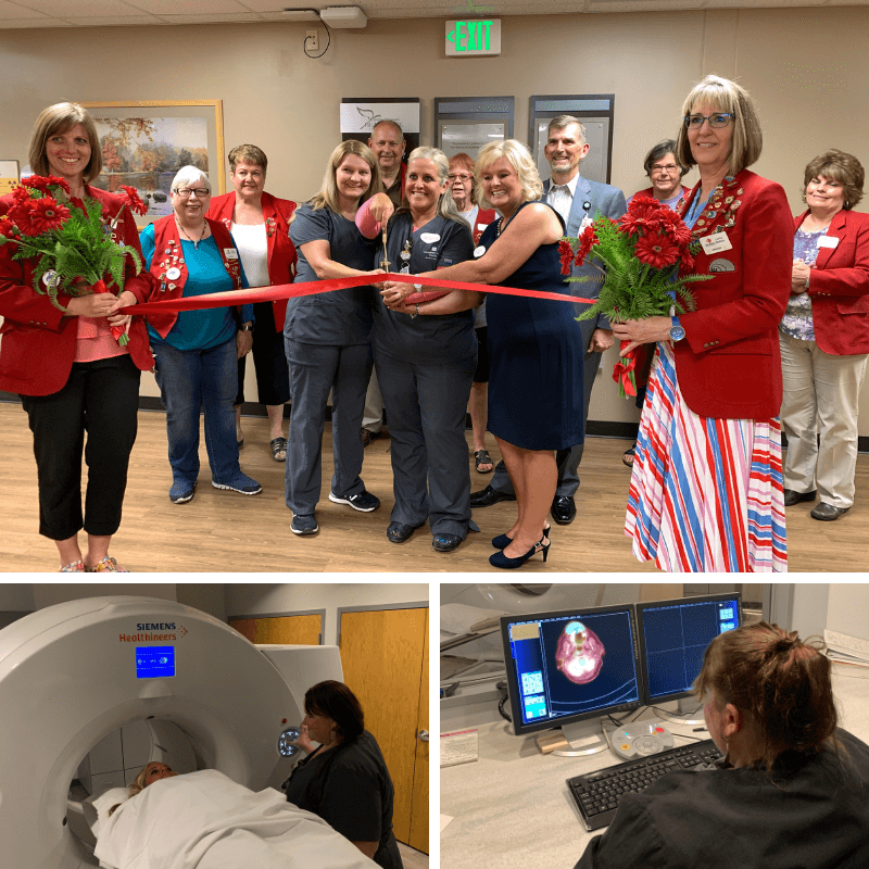
The
Heptner Cancer Center’s new
PET CT is approved and ready for patients. CCH received approval from the
Wyoming Department of Health, and a dedication ceremony was held August 14 to acquaint donors and the
public with the new equipment.
For those of you who don’t know, Positron Emission Tomography (PET)
is a type of nuclear medicine procedure that measures metabolic activity
of the cells of body tissues. PET works by using a scanning device (a
machine with a large hole at its center) to detect photons (subatomic
particles) emitted by a radionuclide in the organ or tissue being examined.
The radionuclides used in PET scans are made by attaching a radioactive
atom to chemical substances that are used naturally by the particular
organ or tissue during its metabolic process. The radionuclide is administered
into a vein through an IV. Next, the PET scanner slowly moves over the
part of the body being examined and uses the information to create an
image map of the organ or tissue being studied. The amount of the radionuclide
collected in the tissue affects how brightly the tissue appears on the
image, and indicates the level of organ or tissue function.
In general, PET scans are used to evaluate organs and/or tissues for the
presence of disease or other conditions. The most common use of PET is
in the detection of cancer and the evaluation of cancer treatment.
More specific reasons for PET scans in cancer treatment include:
- To detect the spread of cancer to other parts of the body from the original
cancer site
- To evaluate the effectiveness of cancer treatment
- To detect recurrence of tumors earlier than with other diagnostic modalities
Newer technology combines PET and CT into one scanner, known as PET CT,
which is what CCH now has. The CT component is used to plan radiation
therapy treatments, according to Cancer Center Director
Leigh Worsley.
“The CT portion of the scanner will be used weekly to scan patients
undergoing radiation therapy treatments for their cancer,” said
Leigh. “The scanner we’ve been using was almost 17 years old
and had a very small opening or ‘bore.’ The new PET CT has
a larger bore and higher quality images that are better for visualization
of tumors and areas of focus. We will also be able to utilize PET CT for
radiation therapy mapping as they are ‘fused’ together and
we will be able to see all metabolic cancer cells that may not be detected
on a traditional CT scan or image.”
PET CT was a joint fundraising project with the Campbell County Healthcare
Foundation and CCH, both contributing nearly $800,000 toward the cost
of the equipment. CCH also funded some structural modifications needed
to house the PET CT equipment.
“I’m so grateful for the generosity of the community and the
support of CCH Administration and Board in this venture,” said Leigh.
“We are excited to be able to provide state-of-the-art cancer imaging
equipment and radiation treatments right here at home!”
Learn more at
www.cchwyo.org/petct.